We provide information sheets on surgical procedures that explain the details of your surgery, written in simple language and illustrated to allow you to better understand what you could expect. The files are in Adobe PDF format available via the links below:
Operations
A-H
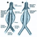
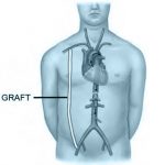
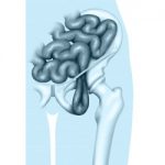
The stent graft is a self-expanding metallic device lined by a tough plastic fabric. It comes in two components: One is introduced through the groin via the iliac artery (the main artery going to the leg) and the top end placed into the aorta above the aneurysm. The bottom end sits in the iliac artery below the aneurysm…
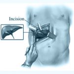
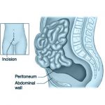
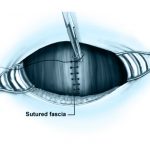
The operation is performed through an incision in the wall of the abdomen. This may be either straight down the midline skirting the umbilicus, from side to side below the umbilicus, or from the left loin to the midline below the umbilicus depending upon the type and extent of the aneurysm that you have…
The leg is amputated through the knee joint. But the skin is much longer to allow it to heal in a wound behind the end of the thigh bone. This operation is only done if the foot is already dead or has lost too much tissue to still be functional or if there is an infection that is too severe to control with antibiotics…
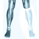
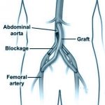
The main artery, which carries blood down your leg, is blocked . The calf, foot and toes are starved of blood. This causes pain on walking. If you have pain at rest or non healing wounds or gangrene this may be a sign of danger to the survival of you leg. We hope to open the blocked part of the artery with a balloon/stent so that blood flows properly again…
The operation is performed through an incision in the wall of the abdomen. This may be either straight down the midline skirting the umbilicus, from side to side below the umbilicus, or from the left loin to the midline below the umbilicus. The choice of incision will be explained by your surgeon during the consultation before arranging the operation…
The operation is performed through an incision in the wall of the abdomen. This may be either straight down the midline skirting the umbilicus, from side to side below the umbilicus, or from the left loin to the midline below the umbilicus. The choice of incision will be explained by your surgeon during the consultation before arranging the operation…
A bowel fistula is an abnormal communication between the small or large bowel or both and the skin. Its causes are multiple and your surgeon will discuss this with you. The principle of the operation is to remove the fistula and a segment of bowel and anastomose (join) the ends of the bowel together…
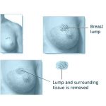
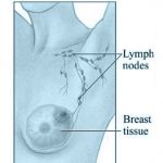
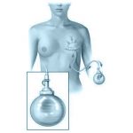
This entails removing the breast lump or an abnormal area on a mammogram which is of concern. This is commonly indicated when there is a breast lump, an area of thickening on the breast or an abnormal area on a mammogram. The removed sample is sent for microscopic examination. Depending on this microscopic examination, further operations may be necessary…
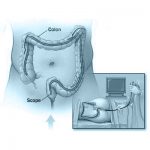
A colonoscopy is a day-case procedure in which the inside of the large intestine, (colon and rectum) is examined using a 1.5 metre flexible instrument with a diameter of less than 1cm. A colonoscopy is commonly used to evaluate gastro-intestinal symptoms, such as, rectal and intestinal bleeding or changes in bowel habit or anaemia…
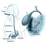
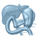
Your symptoms, blood tests and scans point to your gallbladder and bile ducts or pancreas being the cause of your symptoms. ERCP involves passing a flexible telescope (duodenoscope) through the mouth into and through the stomach to the first part of the bowel called the duodenum. Looking through this scope the outlet of the ducts (pipes) from the liver and pancreas can be seen…
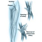
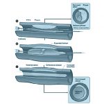
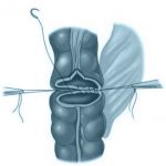
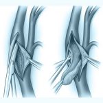
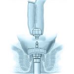
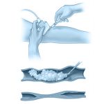
In this operation the plaque of cholesterol material that is causing the blockage in the main artery in the groin is removed altogether by making an incision in the groin. Once the artery has been controlled so that no blood flows through it during the procedure it is opened and the plaque is removed. The artery is then closed with a fabric patch to widen it. No bypass is necessary…
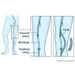
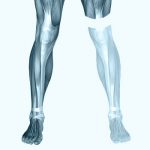
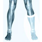
The large arteries in the groin and either above or below the knee are exposed through small incisions. A tunnel is made deep in the leg between these two incisions, and a new bypass graft, usually your own vein but sometimes synthetic material, is passes through the tunnel. The ends of the bypass are then joined to the arteries, so bringing blood from the groin to the lower leg where it is required…
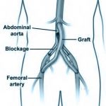
The large arteries in both groins are exposed through small incisions. A tunnel is made under the skin above the pubic bone between these two incisions, and a new bypass graft, usually synthetic material but sometimes your own vein, is passes through the tunnel. The ends of the bypass are then joined to the arteries, so bringing blood from one side to the other where it is required…
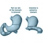
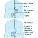
A gastrectomy is an operation to remove part of the stomach (partial) or the entire stomach (total). The small bowel is then joined to the remaining stomach or to the oesophagus. Your surgeon will discuss the procedure with you in detail and will answer any questions you may have before the operation…
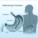
A gastroscopy is an endoscopic examination of your stomach. On the way we examine your oesophagus (gullet, food-pipe) which runs from the back of the mouth, through your chest, and into your stomach. We also examine beyond your stomach into the first part of the gut called the duodenum (part of the small bowel)…
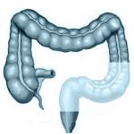
A large bowel resection is an operation to remove all or part of the large bowel or colon because it is diseased or not working properly. The bowel is like a hollow tube. The surgeon will cut out part of the bowel and sew or staple the remaining ends together (an anastomosis). The amount of bowel removed can vary a lot, depending on the reasons for the operation…
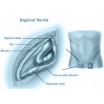
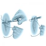
A hernia is a bulge or weakness in the muscles of the stomach in the groin region. There is a weak spot on either side where the arteries and veins run in a tunnel down to your legs and in the case of a hernia some tissue from the inside of your abdomen will protrude along this canal to form a sac that bulge in the groin…
H-Z
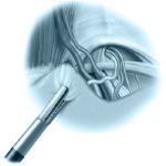
A hernia is a weakness in the muscles which form the lower front of the stomach in the groin region and results in a bulge. This method of hernia repair uses a small telescope under the muscles of the abdomen to examine the weak area. Two other instruments are inserted through the skin and abdomen to dissect away the tissues and display the weakened hernial site. A patch of teflon material is then used to reinforce the weakened area…
A hernia is a bulge or weakness in the muscles which form the lower front of the stomach in the groin region. A cut is made into the skin overlying the hernia. The bulge is pushed back and the sac is cut off. The weak part is mended and strengthened, usually with nylon stitches or a mesh patch which is used to strengthen the defect. The cut in the skin is then closed up…
This is a weakness or swelling of the navel (belly button). It will get bigger and become unsightly. Sometimes fat or bowel gets stuck in the hernia causing severe pain and illness. A cut is made around the navel. Any fat or bowel in the hernia is pushed back or removed. The weakness is mended with strong stitches or a prosthetic sheet (mesh). The skin is then closed up…
During this procedure the colon is removed including the rectum. A new rectum is fashioned from the healthy small intestine and joined internally to the anus to form a ‘pouch’ or reservoir. The surgeon uses part of the healthy small intestine to form the pouch, or reservoir, inside the body and this is attached to the anus. The waste matter from the small intestine goes into this pouch and is passed in the usual manner…
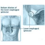
The Nissen fundoplication operation is performed to relieve gastro-oesophageal reflux (GORD). Reflux may be associated with an hiatus hernia. A hiatus simply means a gap/opening . A hernia is a bulge or a weakness. In this case, the stomach bulges through a hiatus up into your chest. Sometimes the stomach gets stuck within the chest. Your surgeon will discuss the procedure with you in detail and will answer any questions you may have before the operation…
This is done when there is a narrowing in the oesophagus(food pipe). It is usually done as an office procedure using a flexible fibre – optic scope but occasionally is done under general anaesthetic. This operation is necessary to facilitate swallowing. The commonest causes are longstanding untreated reflux, a cancer or previous operation to the oesophagus…
An oeso -gastrectomy is a major operation to remove part of the stomach and the lower end of the oesophagus and the remaining stomach is then anastomosed (joined) to the oesophagus. This could be done entirely through the abdomen or combined abdominal and thoracic (chest) – thoraco– abdomina approach, or through the abdomen and chest – transhiatal oesophago – gastrectomy…
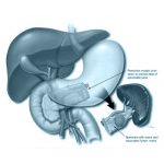
An incision (cut) is made across the upper abdomen (tummy) below the ribs on both sides. The pancreas is exposed and freed from the adjacent organs. The tail (distal) half of the pancreas is then removed. Since the blood vessels that go to the spleen pass through the pancreas, the spleen is also removed…
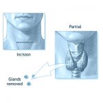
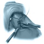
The parotid gland makes saliva to wet the food in your mouth. It is located in your cheek in front of the ear and can extend down to the side of your jawbone. It has a superficial part just under the skin and a deep part that lies deeper on the cheek muscle. The saliva runs from the gland along a tube, which opens into your mouth on either side…
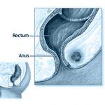
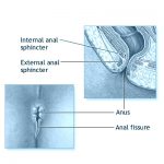
An anal fissure is a small tear running from the skin near the back passage and opening into the anal canal higher up. This is a painful condition usually associated with fresh bleeding after bowel actions. The fissure is aggravated by spasm of the circular anal muscle. This is stretched slightly and some of the muscle fibres divided. This results in relief of the spasm and thus the pain, allowing the fissure to heal. Any associated anal skin tags are removed…
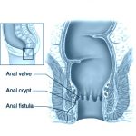
An anal fistula is a narrow tunnel or tract running from the skin near the back passage and opening into the anal canal higher up. This discharges pus irregularly and may be uncomfortable. It often shows up after there has been an abscess near the anus. During the operation the fistula tract is identified and is opened along its length, and the exposed inside of the tract is then cleaned out…
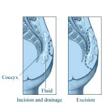
A pilonidal sinus is a condition caused by small hairs collecting under the skin between the buttocks. It is treated by removing the affected skin and involved tissue and closing the resultant defect with stitches. If there is associated infection or an abscess it may be dangerous to close the wound and it is then first drained by a simple operation and then later completely excised once the sepsis has settled…
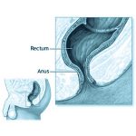
You and your surgeon have decided that your rectal prolapse is severe enough or troublesome enough to need an operation. This operation aims to repair the prolapsed by hitching up the rectum and stitching it to the inside of the pelvis so that it cannot prolapse out through the anus any more. Usually a nylon sheet is placed on the rectum to help secure it. Sometimes the section of bowel above the rectum may have become very slack and elongated and may require to be removed . This procedure is often done laparoscopically (key-hole surgery)…
You and your surgeon have decided that your rectal prolapse is severe enough or troublesome enough to need an operation. A Delorme’s procedure aims to repair the prolapse. This operation involves the surgeon removing some of the prolapsed lining of the rectum (mucosa) and reinforcing the muscle of the rectum by placating stitches. This is done via the anus. No external incision is needed…
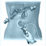
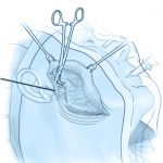
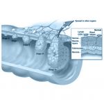
The operation is carried out to remove the spleen. Your surgeon will discuss the procedure with you in detail and will answer any questions you may have before the operation. A telescope with an attached miniature TV camera (laparoscope) is inserted through a small (1 cm) incision above the belly button (umbilicus). Four other similar incisions are made to insert the necessary instruments for the operation and to remove the spleen…
The operation is carried out to remove the spleen.Your surgeon will discuss the procedure with you in detail and will answer any questions you may have before the operation. It is carried out under general anaesthetic and your anaesthetist may place an eoidural for pain control. An incision is made either in the midline down to your umbilicus (belly button) or along the left costal (rib) margin…
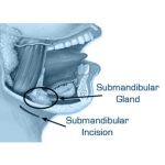
The submandibular gland makes saliva to wet the food in your mouth. It fits under the side of your jawbone. The saliva runs from the gland along a tube, which opens into your mouth under the base of your tongue. If a swelling or stone grows in the gland, the gland has to be removed. If a stone forms in the tube (duct), the stone can usually be removed without taking out the gland…
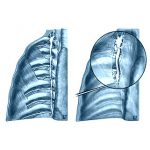
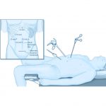
Thoracoscopic Sympathectomy: If the sympathetic nerves to the hands and armpit are to be divided, this is usually done using keyhole surgery to divide the sympathetic chain in the chest. This is done by making 2 small incisions in the armpit and then collapsing the lung under controlled conditions and inserting a telescope into the chest to allow vision of all of the structures in the chest…
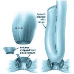
This operation uses a newly developed operating viewing tube (Endoscopic proctoscope) which allows excellent access to the rectum. The rectum is very difficult to work within, because of the limited access via the anus. A colonoscope is usually limited to performing less difficult procedures like biopsy or snaring a simpler polyp…
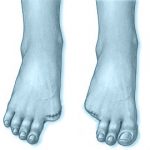
A toenail can become very thick, curved and painful or the nail curls over and grows into the toe, most commonly affecting the big toe but it may happen with other toes as well. Surgical treatment involves either a wedge excision where a part of the nail is removed or it can involve removing the whole nail…
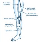
Varicose vein surgery is designed to remove the visible or troublesome varicose veins, and remove the cause of such veins. This entails tying off the superficial vein feeding the varicosities from the deep Veins, and removing the connection between that point and the varicosities themselves. Depending on the type of Varicose Veins you have, either a small incision in the groin or behind the knee will be used to ligate the faulty vein…
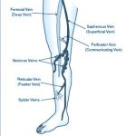
A varicose vein is a skin vein that has swollen because of overspill of blood from veins running deep in the muscles of the legs. Varicose veins cause symptoms and the legs are better without them. There are plenty of deep veins to keep the circulation going. If left, varicose veins can bleed or clot or cause ulcers…
A dilated tortuous superficial vein that is cosmetically unsightly and can give symptoms. Small ones are called spider veins or telangiectasia. SCLEROTHERAPY is the use of an irritant solution to destroy a vein. The solution is placed into the vein by injection to irritate the linings of the vein so causing the walls to become adherent, to collapse and to become fibrosed…
This is a major operation to remove the head of the pancreas. A large incision (cut) is made right across the upper abdomen (tummy) below the ribs on both sides. The abdomen is checked to make sure there are no signs of tumour spread beyond the pancreas. The head half of the pancreas is then cut out, leaving behind the tail half of the pancreas…
Conditions
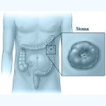
If you suffer from chronic problems with a condition such as ulcerative colitis or familial adenomatous polyposis (F.A.P.), you may be advised that you must undergo surgery to remove the diseased bowel. This will usually mean having an ileostomy – the end of the small intestine is brought to the surface on the abdomen and a ‘stoma appliance bag’ is worn. Many thousands of people have ileostomies and have found that after the operation they enjoy a new improved quality of life -relieved of a constant preoccupation with bowel problems and general poor health…
The most common cause of anal irritation is inappropriate anal hygiene by the patient. Many documents will blame seepage of faeces and moisture and anal hair. While these may contribute in some patients, they are not, in fact, the primary cause in the majority. Other conditions may contribute, but they are all rare. They include allergies, diabetes, inflammatory bowel disease, worms, eczema and psoriasis and fungal infections. Other causes include fleshy anal skin tags, anal fissure and prolapsing haemorrhoids, however the most common cause is assiduous, over-cleansing of the anus…